
Figure 3.
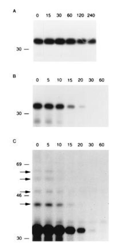
The Met-NP-M1 and Arg-NP-M1 fusion proteins follow the N-end rule. 721.45 cells were infected with Vac-Ub-Met-NP-M1 (A) or Vac-Ub-Arg-NP-M1 (B and C). After [35S]methionine labeling for 10 min (A) or 5 min (B and C), cells were chased for the indicated times in min. Immunoprecipitations of NP-M1 fusion proteins were revealed by SDS/PAGE and autoradiography. Exposure was 6 h in A and 3 days in (B). C is a longer exposure (11 days) of B. Arrows indicate slower migrating bands in C that were not visible in A even after a long exposure. The position of [14C]-labeled markers is indicated on the left.