
Figure 7.
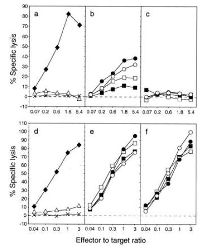
A long half-life is required for the presentation of M1 to HLA-DR1-restricted T cells but not to HLA-A2-restricted T cells. Lysis by the M1-specific HLA-DR1-restricted T-cell clone C3.5 (a–c) or by the M1-specific HLA-A2-restricted T-cell line Q157 (d–f) of 721.45 cells that were untreated (×), incubated with synthetic peptide (♦, M1 18–31 in a, M1 58–66 in d), or infected with Vac-H3 (▵), infected with Vac-Ub-Met-NP-M1 (b and e) or with Vac-Ub-Arg-NP-M1 (c and f). The doses of virus were 30 (○), 10 (•), 1 (□), and 0.3 (▪) pfu/cell and each infection lasted 4 h.