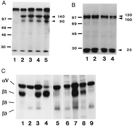
Figure 1.

Immunoprecipitation analysis with the 8-2D, 8-B3, 4-10D, and 8-B11 mAbs. (A) SDS/PAGE analysis of immunopreciptates from lysate of 125I-labeled hybridoma cells run under nonreduced conditions. Lanes: 1, beads alone; 2, 4-10D; 3, 8-B11; 4, 8-2D; 5, 8-B3. Molecular weight standards are indicated in kilodaltons. (B) SDS/PAGE analysis of immunoprecipitates from lysate of 125I-labeled hybridoma cells run under reduced conditions. Lanes: 1, 4-10D; 2, 8-B11; 3, 8-2D; 4, 8-B3. Molecular weight standards are indicated in kilodaltons. (C) SDS/PAGE analysis of immunoprecipitates from lysates of 125I-labeled hybridoma cells and E9 fibroblasts and mixed lysate, run under nonreduced conditions. Lanes: 1, E9 fibroblast lysate, anti-αv serum; 2, hybridoma lysate, anti-αv serum; 3–9, mixed lysate precipitated with: 3, anti-αv serum; 4, anti-β1 serum; 5, anti-β3 serum; 6, 8-2D; 7, 8-B3; 8, 4-10D; 9, 8-B11. Lanes 1–4 were exposed for a shorter time than lanes 5–9, because the anti-αv and anti-β1 sera immunoprecipitate much more efficiently than the other antibodies. T3, hybridoma cells; E9, embryonic d9 fibroblasts; M, mixed lysate.
