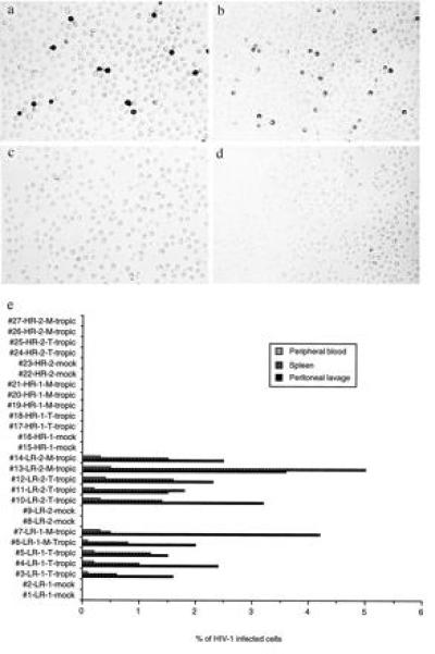
Figure 2.

In vivo infection of hu-PBL-SCID/bg mice. (a and b) Representative HIV-1-positive immunostaining of peritoneal lavage and splenocytes, respectively, of mice reconstituted from a LR individual. (c and d) Representative HIV-1-negative immunostaining of peritoneal lavage and splenocytes, respectively, of mice reconstituted from a HR individual. (e) T cell and macrophage tropic HIV-1 challenge of hu-PBL-SCID/bg mice reconstituted with LR and HR donors’ PBLs. Reconstitution of mice with PBLs from the LR-1 donor (#1–#7), the LR-2 donor (#8–#14), the HR-1 donor (#15–#21), and the HR-2 donor (#22–#27)) were confirmed by ELISA for human Ig and FACS analysis for the human CD45 leukocyte marker. Reconstituted hu-PBL-SCID/bg mice were challenged with 100 TCID50 of T cell (T) or macrophage tropic (M) HIV-1 by i.p. injection. All the mice were killed 4 weeks after reconstitution, and single cell suspensions were prepared from the three different organ compartments for immunostaining. HIV-1-positive cells were counted under an inverted microscope and the percentage of infected cells was determined by taking the average of more than three representative counts of 1000–10,000 cells as previously described (19).
