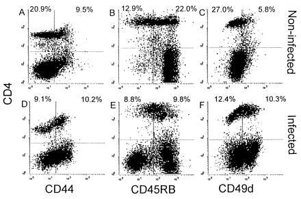
Figure 5.

Activation of splenic CD4+ cells after LCMV infection. Two-color flow cytometric analysis of splenocytes from noninfected β2m− mice (A–C) or from β2m− mice that had been infected i.p. with LCMV 10 days previously (D–F) was performed. Cells were stained for CD4 and CD44 (A and D), CD45RB (B and D), or CD49d (α4-integrin) (E and F). The percentages of CD4+ cells are indicated in the upper two quadrants of each plot.
