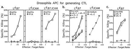
Figure 5.

Generation of primary CTL from naive CD8+ 2C cells cultured with QL9 peptide presented by Drosophila cell APC. (a) CTL activity of CD8+ 2C cells stimulated with Ld.B7, Ld.B7.ICAM, or Ld.ICAM APC plus QL9 peptide (10 μM) in the absence of exogenous cytokines. (b Left) CTL activity of CD8+ 2C cells stimulated with Ld.B7 APC plus QL9 peptide (10 μM) in the absence or presence of anti-IL-2 and/or anti-IL-4 mAb (2 μg/ml). (b Right) CTL activity of CD8+ 2C cells stimulated with Ld.ICAM APC plus QL9 peptide (10 μM) in the absence or presence of recombinant IL-2 (20 unit/ml). (c) CTL activity of CD8+ 2C cells stimulated with Ld.B7 APC plus QL9 peptide in the absence or presence of either anti-IL-2 or anti-IL-4 mAb (20 μg/ml). Purified CD8+ 2C cells (5 × 10⁵) were cultured with 2 × 10⁶ Drosophila APC. After 4 days, the cells were harvested and CTL activity was tested against [51Cr]-labeled RMA-S.Ld target cells loaded with p2Ca or control P1A.35-43 peptide (32); P1A peptide binds strongly to Ld but has no detectable affinity for the 2C TCR (32). The data show the mean level of specific 51Cr release from duplicate cultures.
