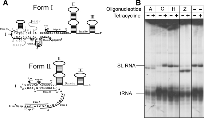
FIG. 5.
Secondary structure of nascently transcribed SL RNA in CBF5-silenced cells. (A) Schematic representation of the secondary structures of the two forms of SL RNA. The secondary-structure predictions are based on data from Harris et al. (11). The three stem-loops and the Sm site of the SL RNA are indicated. The locations of the oligonucleotides (Oligo) used for RNase H cleavage are depicted on the SL RNA structure. The interaction domain of SLA1 with SL RNA is shown in gray. (B) RNase H cleavage of nascently transcribed SL RNA in CBF5-silenced cells. CBF5-silenced cells, uninduced (Tetracycline −) and induced (Tetracycline +) (72 h), were permeabilized and incubated with different oligonucleotides in the presence of RNase H. RNA was extracted from the cells and separated on a denaturing 6% polyacrylamide gel. The designations of the oligonucleotides are shown (their positions are given in panel A). The positions of the SL RNA and tRNA are indicated.

