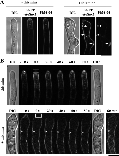
FIG. 6.
Apical recycling and secretory defects in the Aoend4-repressed condition. (A) In the absence (−) of thiamine, EGFP-AoSnc1 was localized mainly in the tip region and was also stained with FM4-64. In contrast, in the presence (+) of thiamine, EGFP-AoSnc1 localization was altered, and it was localized throughout the plasma membrane. FM4-64 accumulated in the large invagination structures labeled with EGFP-AoSnc1 (arrows). The image of a hypha by differential interference contrast (DIC) is shown to the left. Bar, 5 μm. (B) The FRAP analysis was performed in the hyphal tip regions. The time after photobleaching is shown. Photobleaching was carried out in the squares outlined in white of the photographs taken at 0 s; these were taken just after photobleaching. DIC images were taken before (left) and after (right) photobleaching, and these revealed that there was no damage to the hyphae by laser irradiation. The fluorescence recovery of EGFP-AoSnc1 in the hyphal tip was observed in the presence of thiamine at 60 min after photobleaching. Bar, 5 μm.

