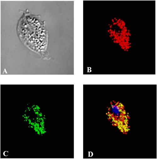
FIG. 4.
Immunodetection of TvFDP in T. vaginalis cells. (A) Nomarski differential contrast; (B) visualization of malic enzyme, hydrogenosomal marker; (C) TvFDP labeling; (D) merge of color channels showing the presence of TvFDP in the hydrogenosomes with DAPI (4′,6′-diamidino-2-phenylindole) staining for nuclei.

