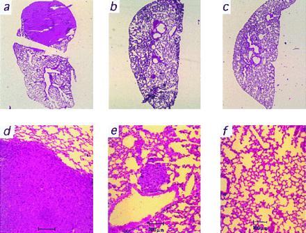
Figure 4.

Representative microscopic analysis of lung metastases growing from melanoma cells producing different amounts of PTN mRNA. Hematoxylin/eosin-stained whole mount sections (a–c) and microscopic views (d–f) of representative lungs from the groups producing high (a and d), medium (b and e), and low levels (c and f) of PTN are shown. For quantitative data, see Fig. 2d and Results.
