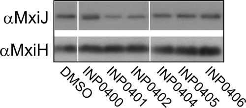
FIG. 5.
Whole-cell levels of T3SS component proteins. Wild-type Shigella cultures were grown in the presence of DMSO or compounds, and then whole-cell extracts were isolated. Samples were separated by SDS-PAGE and analyzed by Western blotting. The antibodies used for the blots are indicated on the left. Lanes were removed for consistency of display of the samples. For each blot, the same exposure was used to make the composite image, and the remaining lanes are shown in their initial loading order.

