
Figure 2.
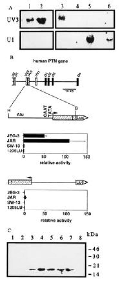
Expression of the PTN gene in various human cell lines and promoter activity of the HERV insert. (A) Northern blot analysis of total RNA from choriocarcinoma cells JEG-3 and JAR (lanes 1 and 2), a chorion biopsy from a stillborn fetus (lane 3), adrenal carcinoma SW-13 (lane 4), melanoma 1205LU (lane 5), and teratocarcinoma PA-1 cells (lane 6) using exon-specific probes. (B) Transcriptional activity of a HindIII–BamHI (H, B) genomic fragment inserted into the promoterless pXP-1 reporter vector (25) in sense and antisense orientation. Data are the mean ± SD of triplicate determinations and are representative of at least two transient transfection experiments for each cell line. Luciferase activity, normalized to the protein content, is expressed relative to that obtained with the pXP-1 vector. Western blot for PTN present in the culture medium of JEG-3 cells. Proteins present in medium conditioned by JEG-3 cells were concentrated and partially purified by heparin-affinity chromatography using a NaCl step gradient of 0.9 to 1.5 M (lanes 1–8) and analyzed as described (4).