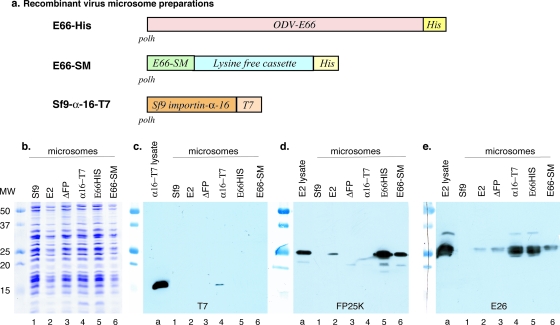
FIG. 4.
Association of E26, FP25K, and importin-α-16 with enriched ER membranes. (a) Schematic of the recombinant viruses used for infection and the preparation of in vitro translation-competent microsomal membranes. In every case, the recombinant gene was inserted into the polyhedrin gene locus under the control of the polyhedrin promoter. (b) Coomassie blue-stained SDS-PAGE gel showing that the microsomal preparations were loaded at approximately equal concentrations. (c to e) SDS-PAGE-separated gels were blotted and analyzed using antibody to T7 epitope (c), FP25K antibody (d), and E26 antibody (e). In panels c to e, lane a shows the control from a total cell lysate obtained from cells infected with the relevant virus. Lanes 1 to 6 represent microsomal membranes prepared from control Sf9 cells (lane 1) and from cells infected with wild-type AcMNPV (lane 2), ΔFP25K virus (lane 3), importin-α-16-T7 virus (lane 4), E66HIS virus (lane 5), and E66-SM virus (lane 6).

