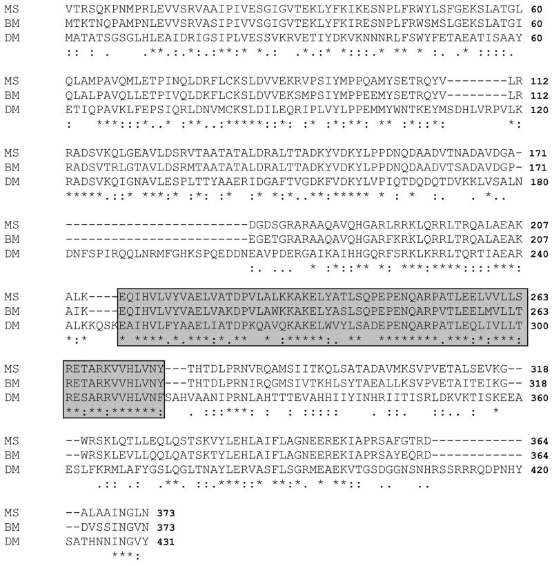
Figure 2. Alignment of Lsd1 deduced amino acid sequences of M. sexta, B. mori and D. melanogaster.
MS (M. sexta, accession number EU809925), BM (B. mori, accession number NP_001040143) and DM (D. melanogaster, accession number NP_732904). Identical (*) and similar (.) residues are indicated underneath the sequences. The alignment was performed using the program CLUSTALW available at www.ch.emb.net.org. The highly conserved region that is predicted to be involved in lipid binding (Arrese et al., 2008) is framed.

