
Figure 6. MsLsd1 transcript levels during development.
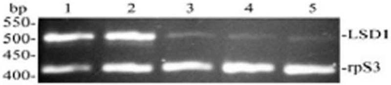
The relative level of the Lsd1 transcript was determined by RT–PCR in a duplex PCR that was performed using primers specific for MsrpS3 (M. sexta ribosomal protein S3) and MsLsd1. Amplification step of the duplex PCR included 22 cycles of 30 s denaturing at 94 °C, 30 s annealing at 56°C, and 1 min extension at 72 °C. The primer concentrations and cycling conditions were optimized in preliminary experiments to avoid saturation. RT-PCR products were separated on 2% agarose gels containing 0.5 μg/ml ethidium bromide and photographed over UV light. A representative gel image is shown. The expected sizes for Lsd1 and rpS3 PCR products are 497 and 415 bp, respectively. Two independent sets of total RNA were independently analyzed at least by duplicate.