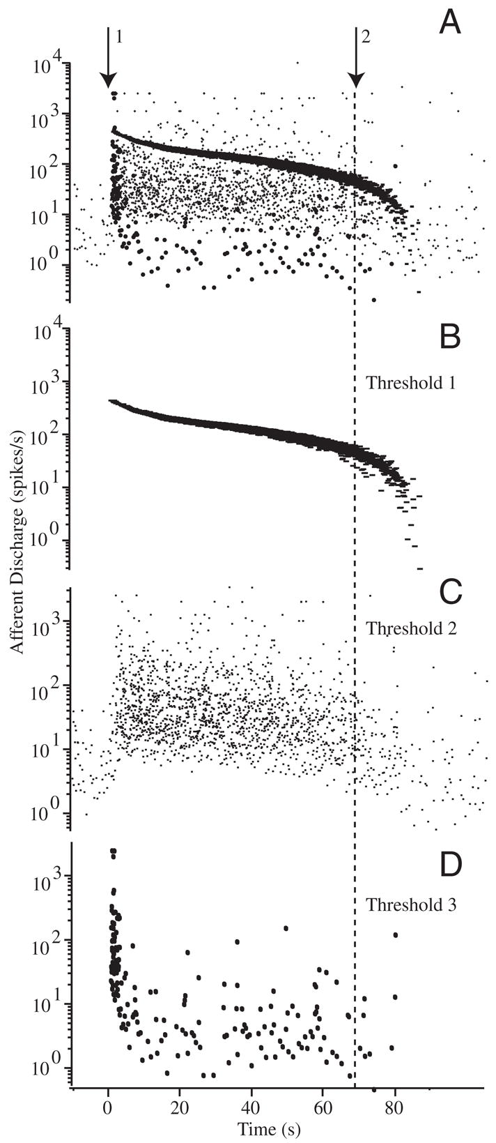
FIG. 5.

Raw afferent responses to canalithiasis: example raw data illustrating the diversity of afferent responses to canalithiasis bead movement. Afferent discharge patterns shown were recorded simultaneously in response to a single epoch of bead movement in an example animal. A: extracellular action potentials recorded using glass suction electrode attached to the side of the lateral canal nerve branch. Arrow 1 indicates the instance when beads were introduced into the LC and arrow 2 indicates when the beads stopped moving. Window discrimination was applied to associate voltage spikes with individual afferent units. B and C: examples of 2 different afferent units exhibiting tonic increases in discharge over the entire duration of bead movement. Discharge rate returned to the prestimulus background rate only after the beads completely stopped moving (lasting >60 s). D: example of a third afferent exhibiting and initial increase in discharge rate followed by a period of adaptation that returned the discharge to the prestimulus rate before the beads stopped moving.
