
Fig. 7.
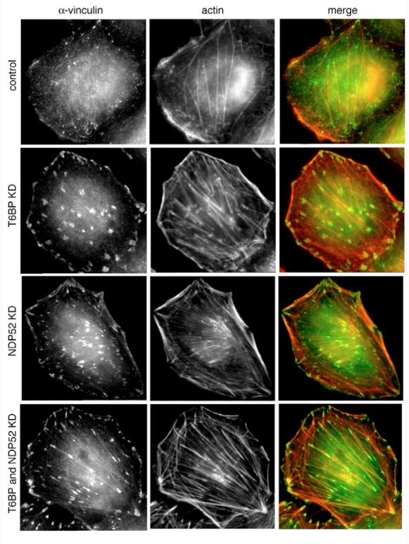
Loss of T6BP and NDP52 leads to dramatic changes in actin filament organisation and focal adhesion formation. HeLa cells were depleted of T6BP, NDP52 or both proteins by transfection with siRNA and stained for immunofluorescence microscopy using Rhodamine-phalloidin and antibodies against vinculin. Whereas control cells show membrane ruffles at the plasma membrane but few focal adhesions and stress fibres, the KD cells have lost membrane ruffles and display a substantial increase in size and number of focal adhesions and stress fibres, respectively.