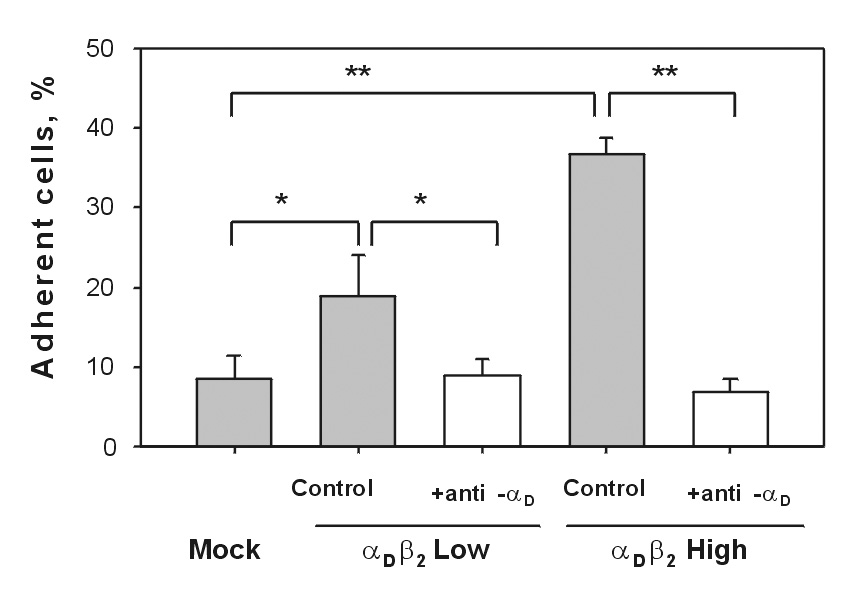
Fig.2. Adhesion of HEK 293 cells expressing different densities of αDβ2 and mock-transfected HEK 293 cells to vitronectin.

Aliquots of calcein-labeled cells (5×10⁴) were allowed to adhere to wells of 96-well plates coated with 5 µg/ml vitronectin in the absence or presence of blocking anti-αDβ2 antibody (10 µg/ml). After incubation for 30 min at 37°C in 5% CO2, the nonadherent cells were removed by two washes with PBS and fluorescence was measured. The number of adherent cells was calculated by using the fluorescence of aliquots with a known number of labeled cells. Data are expressed as a percentage of added cells and are the mean ± SD of three individual experiments (* denotes P<0.05; ** denotes P<0.01).
