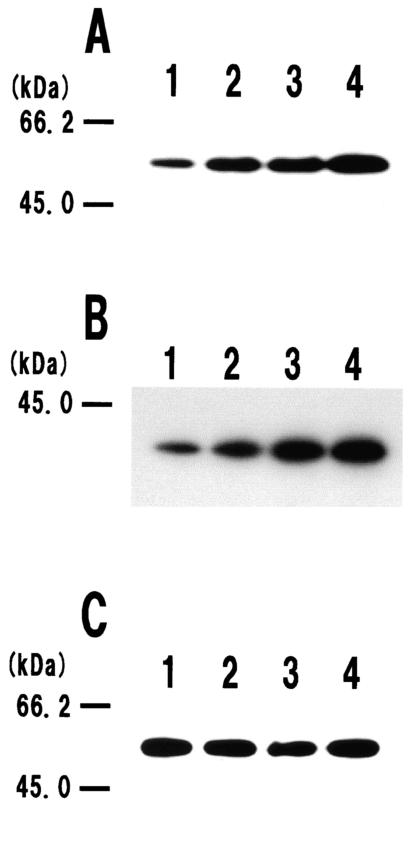
FIG.1.
Effects of extracellular proteases on degradation of the LytF-3xFLAG (A), LytE-3xFLAG (B), and LytC-3xFLAG (C) fusion proteins. Electrophoresis was performed on SDS-12% polyacrylamide gels. Cell surface proteins were prepared and subjected to Western blot analysis as described in Materials and Methods. The molecular masses of the protein standards (Bio-Rad) are indicated on the left. We confirmed the reproducibility of Western blot analysis in at least three independent experiments. (A) Western blot of the LytF-3xFLAG fusion protein. Lanes 1 to 4 contained cell surface extracts of the E3FL (lytF-3xFLAG), EPRE3FL (epr lytF-3xFLAG), WAE3FL (wprA lytF-3xFLAG), and WE1E3FL (wprA epr lytF-3xFLAG) strains, respectively. Cells were cultured in LB medium at 37°C and were harvested at the transition stage (OD600, 1.9). Equal amounts of cell surface proteins (equivalent to 0.15 OD600 unit) were applied to the lanes. (B) Western blot of the LytE-3xFLAG fusion protein. Lanes 1 to 4 contained cell surface extracts of the F3FL (lytE-3xFLAG), EPRF3FL (epr lytE-3xFLAG), WAF3FL (wprA lytE-3xFLAG), and WE1F3FL (wprA epr lytE-3xFLAG) strains, respectively. Cells were cultured in LB medium at 37°C and were harvested at the transition stage (OD600, 1.8). Equal amounts of cell surface proteins (equivalent to 0.1 OD600 unit) were applied to the lanes. (C) Western blot of the LytC-3xFLAG fusion protein. Lanes 1 to 4 contained cell surface extracts of the B3FL (lytC-3xFLAG), EPRB3FL (epr lytC-3xFLAG), WAB3FL (wprA lytC-3xFLAG), and WE1B3FL (wprA epr lytC-3xFLAG) strains, respectively. Cells were cultured in LB medium at 37°C and were harvested at the transition stage (OD600, 2.3). Equal amounts of cell surface proteins (equivalent to 0.1 OD600 unit) were applied to the lanes.

