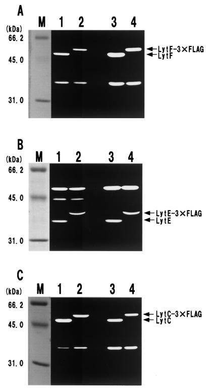
FIG.2.
Zymography of the 3xFLAG fusion proteins. Cell surface proteins from cells expressing the LytF-3xFLAG (A), LytE-3xFLAG (B), and LytC-3xFLAG (C) fusion proteins were extracted as described in Materials and Methods. Equal amounts of proteins (equivalent to 10 OD600 units) were applied to the lanes. We confirmed the reproducibility of zymography in at least three independent experiments. (A) Detection of the LytF-3xFLAG fusion protein. Cells were cultured in LB medium at 37°C and were harvested at the transition stage (OD600, 2.2). Lane M, size marker; lane 1, wild type; lane 2, E3FL (lytF-3xFLAG); lane 3, WE1 (wprA epr); lane 4, WE1E3FL (wprA epr lytF-3xFLAG). (B) Detection of the LytE-3xFLAG fusion protein. Cells were cultured in LB medium at 37°C and were harvested at the transition stage (OD600, 2.0). Lane M, size marker; lane 1, wild type; lane 2, F3FL (lytE-3xFLAG); lane 3, WE1 (wprA epr); lane 4, WE1F3FL (wprA epr lytE-3xFLAG). (C) Detection of the LytC-3xFLAG fusion protein. A lytF mutation was introduced into each strain used for zymography since LytF and LytC overlapped, as described in Materials and Methods. Cells were cultured in LB medium at 37°C and were harvested at the transition stage (OD600, 2.4). Lane M, size marker; lane 1, ED (lytF); lane 2, B3FLEd (lytF lytC-3xFLAG); lane 3, WEEd (wprA epr lytF); lane 4, WEB3FLEd (wprA epr lytF lytC-3xFLAG).

