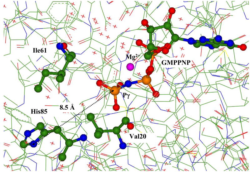
Figure 1.
The fragment of the crystal structure PDBID:1EXM (15) showing the positions of GTP analog, GMPPNP, magnesium cation, His85, and the side chains of the ‘hydrophobic gate’ Val20 and Ile61. Here and below the carbon atoms are distinguished by green, oxygen atoms by red, nitrogen atoms by blue, phosphorus atoms by brown, magnesium by magenta.

