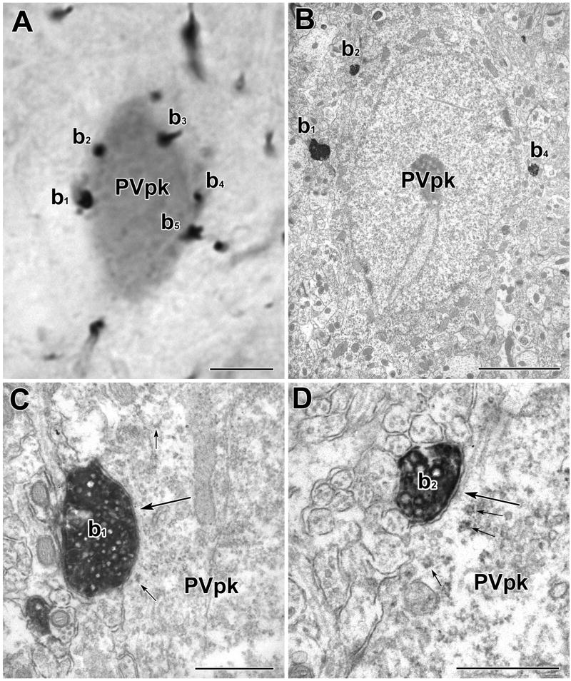
Fig. 10.
A PV+ perikaryon is enveloped by TH+ axons in a basket-like configuration. A) Light micrograph of a PUR-labeled PV+ perikaryon (PVpk) receiving apparent contacts from DAB-labeled TH+ terminals (b1–b5). B) Electron micrograph of the PUR-labeled PV+ perikaryon (PVpk) shown in A. C) Higher power view of the contact (large arrow) formed by TH+ terminal b1. This contact was considered to be a synapse with a narrow synaptic cleft (see Experimental Procedures). Small arrows indicate examples of PUR label. D) Higher power view of the contact (large arrow) formed by TH+ terminal b2. This contact was considered to be a small synapse. There is a pronounced density in the synaptic cleft. A cluster of synaptic vesicles adjacent to the presynaptic membrane was more obvious in an adjacent thin section. Small arrows indicate examples of PUR label. Scale bars = 5 μm in A and B, 500 nm in C and D.

