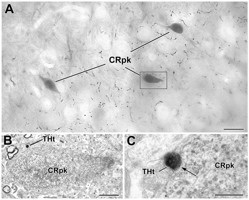
Fig. 5.
A TH+ terminal contacts a CR+ perikaryon in the BLa. A) Low power photomicrograph (grayscale image) of an osmicated, resin−embedded section showing three PUR-immunostained CR+ perikarya (CRpk) in the BLa at the bregma −2.8 level. Most of the varicose axons in the neuropil are TH+. The perikaryon in the box is shown in B. B) Electron micrograph of a CR-immunoreactive perikaryon. The TH+ terminal (THt) contacting the CR+ perikaryon is shown in C. C) Higher power electron micrograph of the DAB-labeled TH+ terminal forming an apposition (arrow) with the PUR-labeled CR+ perikaryon shown in B. Note the particulate PUR reaction product in the CR+ perikarya. Scale bars = 10 μm in A, 2 μm in B and 500 nm in C.

