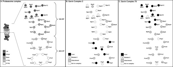
Figure 3.
Two-dimensional structural representation of the proteasome 19S RP and 20S CP, overlaid with high-throughput data from affinity purified protein complexes. (A) 2-D representation as in Figure 1A. (B) Core, module and attachment proteins from Complex 2 [12] overlaid on the 2-D representation. Note that the core proteins are mostly those in the 20S CP. (C) Core, module and attachment proteins from Complex 75 [12] overlaid on the 2-D representation. Note that the core proteins here are mostly those in the proteasome lid and hinge.

