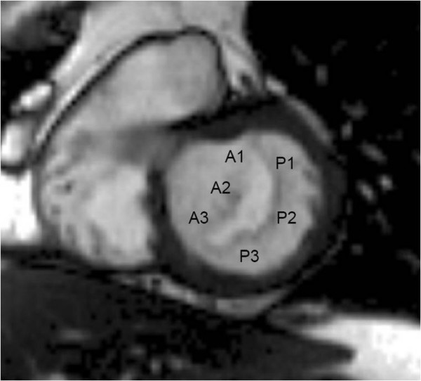
Figure 1.

Anatomy of the mitral valve. CMR short axis view of the mitral valve from a basal short axis slice showing its two leaflets (anterior and posterior) and the three scallops of each leaflet (A1, A2 and A3 in the anterior leaflet, P1, P2 and P3 in the posterior leaflet). The mitral valve is viewed from the LV looking towards the left atrium.
