
Figure 4.
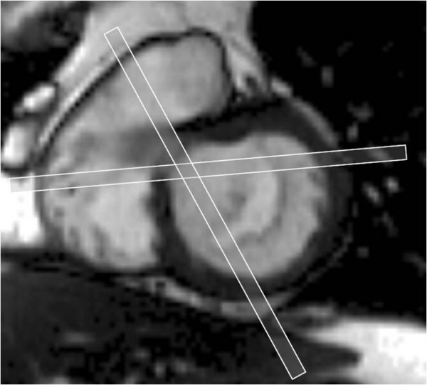
Additional imaging slices at each end of the mitral valve. A further pair of slices orthogonal to the coaptation plane of the valve leaflets is taken at the commissures at each end of the mitral valve (A1-P1 and A3-P3) to better visualise these scallops.