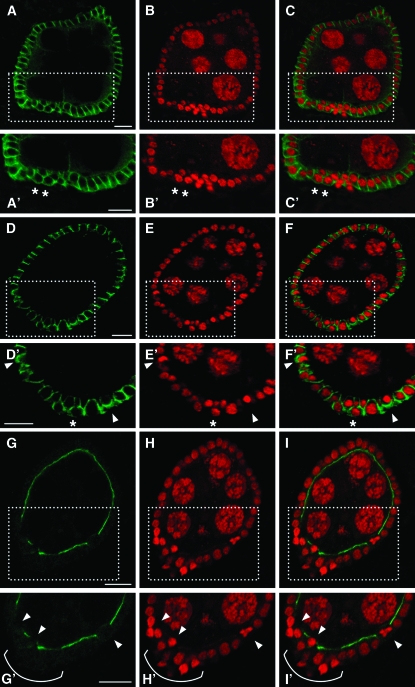
Figure 5.—
Knockdown of EcR-B1 isoform in follicle cells affects apical and basolateral polarity. Confocal cross-section of stage 7 egg chambers from females in which the Cy2-Gal4 driver controls the UAS-IR-EcR-B1 expression (A–I′). Boxed areas in A–I are enlarged in A′–I′, respectively. Propidium iodide staining shows the piling up of follicle cells whose nuclei are strongly stained and show altered shape (B, B′, E, E′, H, and H′; red). Dlg immunodetection (A and A′; green) in multilayered follicle cells (asterisks in A′–C′) shows a circumferential localization of this protein (C′; merged propidium iodide and Dlg signals). The Scrib signal (D and D′; green) is detected at higher levels and it is mislocalized. Follicle cells with strong nuclei staining show a basal Scrib localization (see arrowheads in D′, E′, and in their merge F′). Asterisks in D′–F′ point to a delaminating follicle cell that exhibit apico-lateral distribution of the Scrib signal. Apical aPKC localization (G and G′; green) in multilayered follicle cells (H and H′; red) is absent (I and I′; merged propidium iodide and aPKC signals) (see bracket in G′–I′). Also some follicle cells facing the germline lack apical aPKC staining (see arrowheads in G′–I′). Anterior is up in all panels. Bars, 10 μm.

