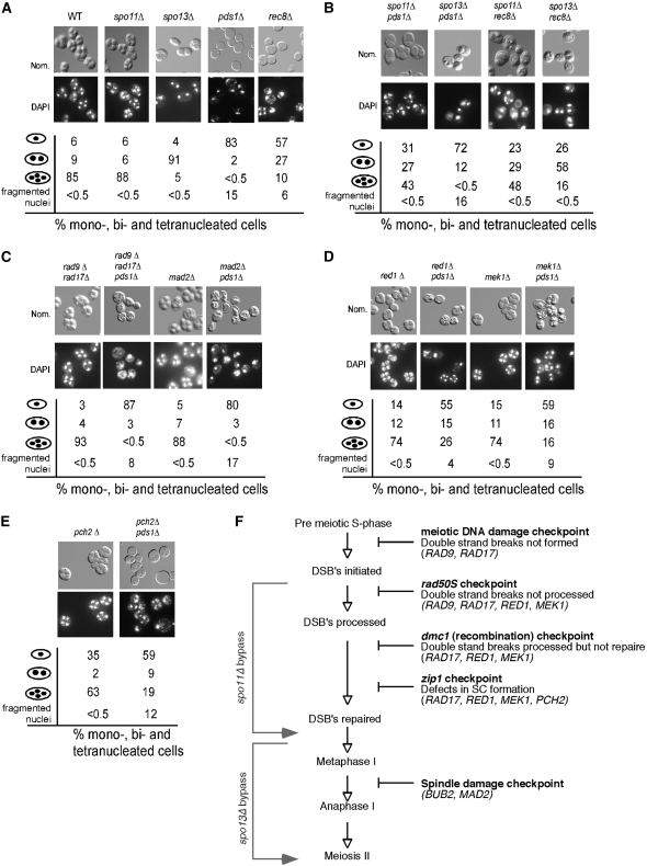
Figure 2.—
Recombination initiation is required for the pds1Δ-induced meiotic arrest. (A) The meiotic progression and spore-wall assembly of wild-type (RSY335), spo11Δ (KCY198), spo13Δ (RSY767), pds1Δ (RSY787), and rec8Δ (KCY385) strains were assayed after 24 hr at 23° by DAPI analysis (bottom) and Nomarski imaging (top), respectively. Population percentages of mono-, bi-, tri-, and tetra-nucleated cells as well as irregular nuclei (scored as fragmented nuclei) in each culture are indicated. (B) Same as in A except that spo11Δ pds1Δ (KCY207), spo13Δ pds1Δ (RSY795), spo11Δ rec8Δ (KCY398), and spo13Δ rec8Δ (KCY399) strains were assayed for nuclear division and spore formation. (C) Same as in A except that rad9Δ rad17Δ (KCY450), rad9Δ rad17Δ pds1Δ (KCY453), mad2Δ (RSY740), and mad2Δ pds1Δ (RSY864) strains were assayed for nuclear division and spore formation. (D) Same as in A except that red1Δ (RSY1355), red1Δ pds1Δ (RSY1358), mek1Δ (RSY1356), and mek1Δ pds1Δ (RSY1359) strains were examined. (E) Same as in A except that pch2Δ (RSY1536) and pch2Δ pds1Δ (RSY1537) strains were examined. For all morphology quantitations presented, the standard deviations were ≤9% for all values. Magnification is ×1000. All the strains used are isogenic to RSY335, the SK1/W303 parent. (F) Diagram depicting the meiotic checkpoints tested with the bypass function of the genes assayed indicated.

