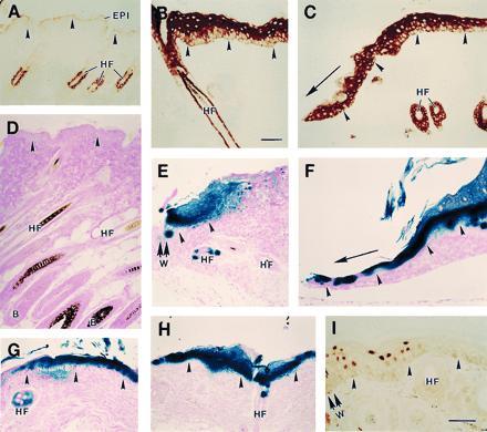
Figure 2.

Inducible expression of lacZ transgene in transgenic mouse skin. (A–C) Cross-sections of control mouse skin reacted with an anti-mouse K6 antiserum followed by a peroxidase conjugate. Shown are intact skin (A), PMA-treated skin (B), and wound edge tissue (C) at 48 hr after skin injury. (D–H) Cross-sections of skin tissue from LacZ transgenic mice processed for β-gal histochemistry. (D) Intact skin from a [5.2-kb hK6a]-LacZ transgenic mouse. (E and F) Wound edge tissue at 4 hr (E) and 3 days (F) after skin injury in a [5.2-kb hK6a]-LacZ transgenic mouse (W, wound site). (G and H) PMA-treated skin tissue from a [5.2-kb hK6a]-LacZ transgenic mouse (G) and a [2.5 kb hK6a]-LacZ transgenic mouse (H). (I) The wound edge (W) of a [2.5-kb hK6a]-LacZ transgenic mouse at 24 hr after full-thickness skin injury reacted with an anti-β-gal antibody followed by a peroxidase conjugate. In all frames the dermo-epidermal interface is highlighted with arrowheads. In C and F, the direction of migration of the epithelium is depicted with an arrow. EPI, epidermis; HF, hair follicle; B, HF bulb region. (Bars = 100 μm.)
