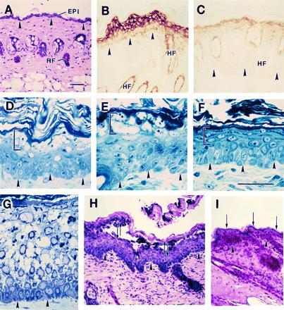
Figure 3.

Induction of mutant keratin expression results in trauma-induced epidermolysis in mouse skin. (A) Hematoxylin/eosin stained section of intact skin in a [5.2-kb hK6a]-ΔK6a-myc mouse (21–1p line). (B) Myc antibody-stained section of paraffin-embedded, PMA-treated skin of a transgenic mouse that expresses the mutant transgene at high level (Fig. 4, lane 6; line 21–1p), whereas C shows myc staining in a mouse that expresses the transgene at low level (Fig. 4, lane 7; line 21–1 m). (D–F) Toluidine blue-stained sections of epoxy-embedded mouse skin. (D and E) PMA-treated skin of 21–1p transgenic mice; (F) PMA-treated control skin. Bracketed areas depict histological aberrations in transgenic epidermis but not in control. (G) Toluidine blue-stained section of epoxy-embedded skin from a individual suffering from severe epidermolytic hyperkeratosis. (H and I) Application of mechanical trauma by repeated tape-stripping in PMA-treated skin of a 21–1p transgenic mouse (H) and a control mouse (I). In H the opposing arrows denote the rupture of epidermis in the uppermost spinous layers, while the arrows in I denote the maintenance of the integrity of the granular layer. Arrowheads highlight the location of the dermo-epidermal interface. EPI, epidermis; HF, hair follicle. (Bars = 100 μm.)
