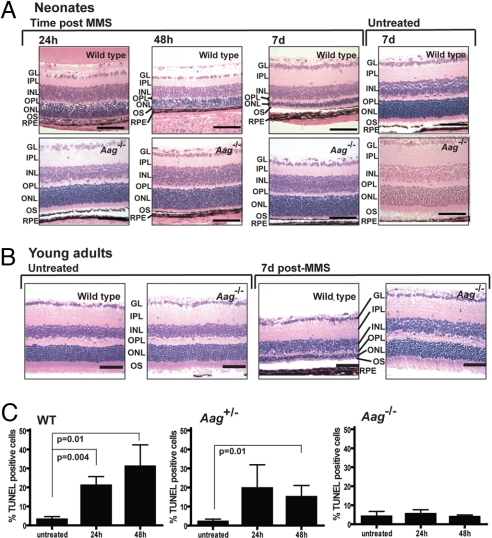
Fig. 2.
Aag-dependent photoreceptor cell death occurs shortly after MMS treatment in both neonates and young adult wild-type animals. (A) Animals were treated at PND7. MMS-induced photoreceptor cell death was seen as early as 24 h after i.p. injection in a wild-type mouse, continued at 48 h after drug, and led to severe degeneration in the wild-type retina 7 days after treatment. Aag−/− animals are completely resistant to MMS, and 7 days after MMS administration there is no evidence of any photoreceptor cell loss in the retinas of Aag−/− animals. GL, ganglion cell layer; IPL, inner plexiform layer; INL, inner nuclear layer; OPL, outer plexiform layer; OS, outer segments. (Magnification: 400×; bar: 100 μm.) (B) Animals treated at 6–8 weeks of age with MMS. Representative photomicrographs are shown at a magnification of 400×. (Bars: 100 μm.) (C) Histogram graph representing quantification of MMS-induced retinal degeneration at the neonatal stage in at least 3 animals per time point. Apoptotic retinal cell death was quantified by flow cytometry in wild-type, Aag+/−, and Aag−/− animals. Error bars represent SD.

