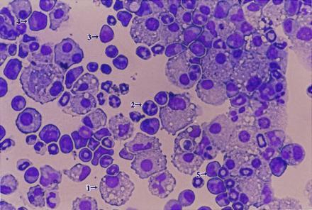
Figure 3.

Analysis of cells found in mixed colonies formed on C166 cells. Cells were removed from mixed colonies grown on C166 feeder layers and evaluated for cell differentiation by Wright–Giemsa staining of cytospin preparations. Representative examples of various cell types are indicated by arrows: 1, macrophage; 2, granulocyte; 3, blast cell; 4, megakaryocyte; and 5, monocyte. (×160.)
