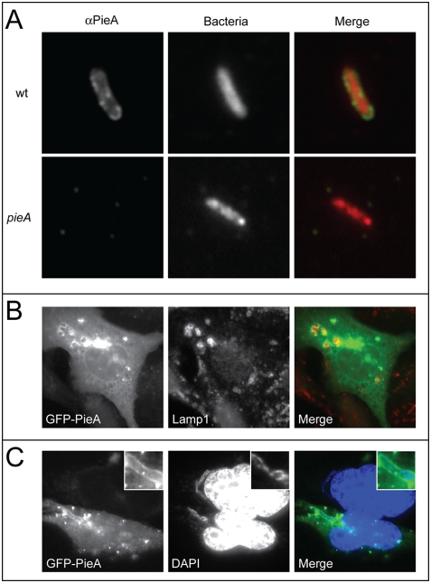
Figure 3. PieA associates with vacuoles containing L. pneumophila.
(A) Representative epifluorescence micrographs of L. pneumophila vacuoles isolated from U937 macrophage-like cells infected with bacteria expressing the fluorescent protein DsRed. Endogenous PieA was detected on vacuoles isolated from cells infected with wild-type L. pneumophila using a PieA-specific antibody and FITC-labeled secondary antibodies. No PieA staining was detected on vacuoles from cells infected with a pieA mutant strain. CHO FcγRII cells transiently expressing the fusion protein GFP-PieA were (B) fixed and stained for the lysosomal marker LAMP-1, revealing GFP-PieA staining in regions of clustered lysosomes, or (C) infected with wild-type L. pneumophila for seven hours, and stained with DAPI to identify bacterial DNA and host cell nuclei. Vacuoles containing L. pneumophila are magnified in the inset of each image. GFP-PieA was observed in association with vacuoles containing L. pneumophila.

