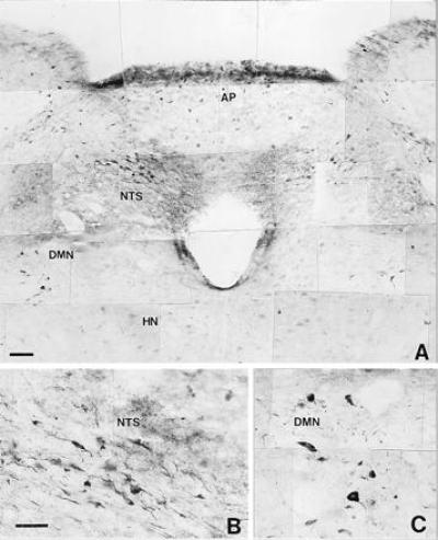
Figure 9.

Microphotograph showing nNOS immunoreactive neural structures distributed in the most rostral portion of the medulla oblongata (A) from a hyperthermic rat (39°C). HN, hypoglossal nucleus. Note that the microphotograph shows an increase in the immunoreactivity in the NTS and DMN. Some neurons of the hypoglossal nucleus show light immunoreactivity. (B and C) Greater magnification of immunoreactive neurons in the NTS and DMN, respectively. (Bars = 0.005 mm.)
