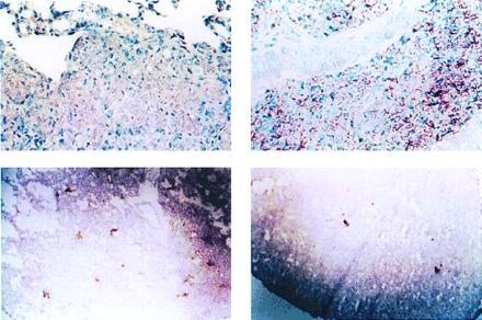
Figure 2.

Immunohistopathological studies of tissues of M. tuberculosis-infected CD1 mice fed a 20% (Left) or 2% (Right) protein diet. Kinyoun’s acid fast stain of lung tissues (Upper) reveals markedly increased bacillary loads in mice with PCM (25 days postinfection) compared with those of controls. Difference in tissue bacillary loads is observed as early as 14 days postinfection in the lungs. Studies of hepatic tissues demonstrated similar findings. Immunocytochemical staining of lung tissues (2.5×) using anti-iNOS antibodies (Lower) demonstrates deficient expression of macrophage iNOS in the lungs of M. tuberculosis-infected mice with PCM. Sections represent tissues obtained from mice 10 days postinfection.
