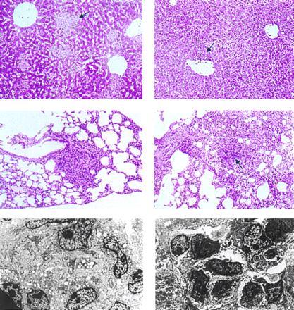
Figure 4.

Granuloma formation in CD1 mice with PCM (Right) is deficient during tuberculous infection (106 cfu M. tuberculosis i.v.) compared with controls given a full protein diet (Left). (Top) Hematoxylin/eosin (H&E)-stained hepatic tissues from mice 7 days postinfection (×20) showing differences in granulomas. In contrast to the well-organized granulomas (arrow) in mice fed a full protein diet (Top Left), those in animals with PCM (Top Right) contain diffuse cellular infiltrates (arrow). (Middle) Necrosis in granulomas in lung tissues of mice with PCM (H&E stained, 14 days postinfection; ×10): Areas of necrosis (arrow) in granulomas of M. tuberculosis-infected mice with PCM (Middle Right). (Bottom) Organization of granulomas: Electron micrographs of liver tissues from mice fed a regular diet (Bottom Left) at 25 days postinfection reveal granulomas with tightly apposed cellular components, compared with those from mice with PCM (Bottom Right), which are loosely organized with apparent intercellular spaces (arrows).
