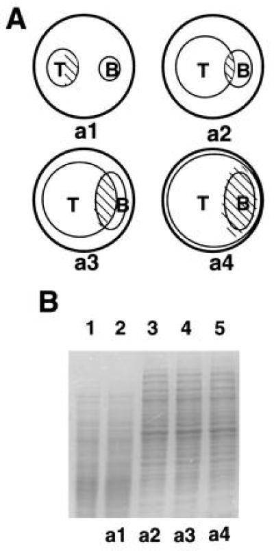
Figure 1.

Plate confrontation assay of T. harzianum and B. cinerea, and SDS/PAGE of cell-free extracts of mycelia collected during different stages of mycoparasitism. (A) Schematic description of the plate assay used to prepare cell-free extracts from different stages of mycoparasitism. Small circles represent T. harzianum (T) or B. cinerea (B) colonies growing on agar medium in a Petri plate (large circles). The mycelium was collected from the pattern-filled zones before the contact (a1), and 12 h (a2), 24 h (a3), and 72 h (a4) after the contact. Samples were also collected from other conditions (see Materials and Methods). (B) SDS/PAGE of T. harzianum cell-free extracts from different conditions. Lanes: 1, extract from a T. harzianum–T. harzianum interaction 12–24 h after the contact; 2, T. harzianum extract from mycelium in stage a1; and 3–5, T. harzianum extracts of mycelia from stages a2, a3, and a4, respectively. Twenty micrograms of protein was applied per slot.
