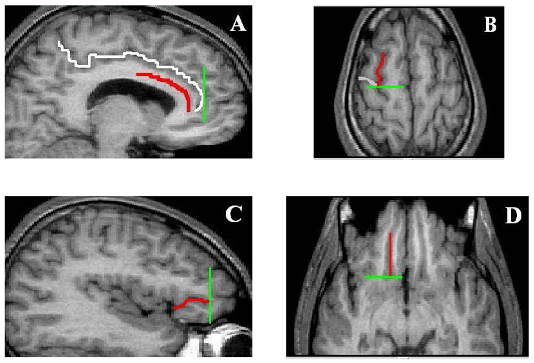
Figure 2.
Illustration of the Limiting Boundaries for the Frontal Lobe Regions
Notes. Panel A. Cingulate sulcus (white), callosal sulcus (red) and tip of the cingulate sulcus (green); Panel B. Superior frontal sulcus (SFS; red), precentral sulcus (PRC; white) and connection of the SFS and PRC (green); Panel C. anterior horizontal ramus (ahr; red) and anterior tip of the ahr (green); Panel D. Olfactory sulcus (OLS, red) and posterior tip of the OLS (green)

