
Figure 1.
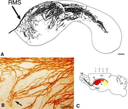
Network of PSA-NCAM immunopositive chains in the lateral wall of the lateral ventricle of adult mice. Dorsal is up and rostral is left. (A) Camera lucida drawing of PSA-NCAM immunopositive chains in whole mounts of this wall. The dorsal group of chains on the wall of the anterior horn are connected to the RMS, but the tissue broke at this point and others (dashed line) during processing. The outline corresponds to the colored area in C. The rectangle indicates the area shown in B. (B) Photomicrograph of PSA-NCAM-immunopositive chains shown in marked rectangle in camera lucida drawing in A. Notice a group of chains (arrow) that are disorganized and end in the central region of the anterior horn. (C) Schematic sagittal view of adult mouse brain showing in different colors the different regions of the lateral wall of the lateral ventricle. The medially located anterior horn (red) is connected to the laterally located inferior horn (yellow) through an intermediate bridge (orange). Black arrow indicates direction of migration of neuronal precursors in the RMS to the olfactory bulb, where cells disperse radially (thin arrows) to reach the granular and glomerular layers (12). Numbers indicate anterior-posterior (A-P) stereotaxic coordinates (measured in mm). OB, olfactory bulb; M, foramen of Monro; NC, neocortex; CB, cerebellum; cc, corpus callosum. (A, bar = 250 μm; B, bar = 100 μm.)