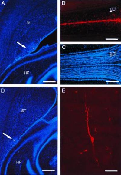
Figure 4.

Microinjection of DiI into caudal SVZ results in labeled cells in the olfactory bulb. (A) DiI microinjection site at A-P −1 (arrow) in the SVZ at the level of the anterior hippocampus (horizontal section). (B) In this same brain, 30 days after injection, many DiI-labeled cells have reached the core of the olfactory bulb (the rostral extension of the RMS) and have migrated into the granule cell layer. (C) Hoechst 33258 counterstaining of section shown in B, to indicate olfactory bulb anatomy. (D) DiI microinjection site at A-P −1.5 (arrow) in the SVZ at the level of the hippocampus. (E) DiI-labeled granule neuron in the olfactory bulb of brain shown in D. HP, hippocampus, ST, striatum, gcl, granule cell layer. (A and D, bar = 450 microns; B and C, bar = 400 microns; bar = 20 microns.)
