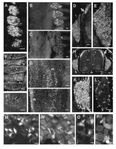
Figure 1.

Darkfield micrographs of DRGs (A–C and F) and spinal cord (G and J) after hybridization with probe complementary to ppGAL mRNA and fluorescence micrographs after incubation with GMAP antiserum (D, E, H, I, K–M, and O). C shows the same section as B stained with bisbenzimide. L shows semiadjacent sections to K incubated with GMAP antiserum preabsorbed with an excess of GMAP. N and P show the same sections as M and O, respectively, after staining with propidium iodide. Distinct ppGAL-mRNA and GMAP expression is seen in DRGs (g) at E15 (A, D, and E), E17 (B, H, and K), and E19 (F). Immunoreactive fibers (arrows) run from the DRGs to the dorsal horn (D, H, and K). GMAP-positive cell bodies are shown in the ventral horn (I). Confocal micrographs show immunoreactivity in mainly the Golgi compartment (arrows) but also as dot-like structures in the thin perinuclear cytoplasm (small arrows) (M–P). The GAL-R1 receptor mRNA is at E17 and is found mainly in the ventral horn (vh) (G) and at E19 in addition in the dorsal horn (dh) (J). Arrowheads in D, H, K, and L point to nonspecific staining, as revealed in the absorption experiments (L). v, vertebra. (A–D, F–H, and J, bar = 100 μm; I, K, and L, bar = 50 μm; E, bar = 25 μm; M and N, bar = 5 μm; O and P, bar = 2.5 μm.)
