Figure 2.

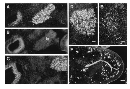
Darkfield (A and C–E) and fluorescence micrographs (F) of inner ear (A and C), trigeminal ganglion (A and C–E), and lip (F) showing ppGAL mRNA (A and C–E) and GMAP-LI (F). B shows propidium iodide counterstaining of sections in A. D and E are semiadjacent sections. ppGAL bmRNA expression is seen in the trigeminal ganglion (tg) at E15 (A) and E17 (C and D). GAL-R1 mRNA is present in apparently fewer cells (E). Also, the sensory epithelium (e) of the inner ear shows GAL mRNA expression (A and C). GMAP-LI can be seen in a nerve extending branches in the skin, including into the epithelium (big arrowheads) (F). Small arrowheads point to nonspecific staining. (A–C, bar = 100 μm; D and E, 50 μm; F, bar = 200 μm.)
