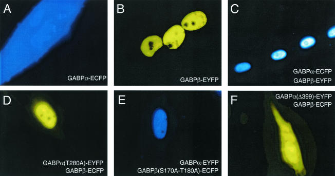
FIG. 2.
Localization of ECFP- or EYFP-tagged GABP subunits in living C2C12 myotubes. Epifluorescence microscope images were taken with the filter set given by the color in the panel. Images were taken 4 to 6 days after induction of differentiation. C2C12 myoblasts were transfected with GABPα-N1-ECFP (A) or GABPβ-C1-EYFP (B) or cotransfected with GABPα-N1-ECFP and GABPβ-C1-EYFP (ECFP filter set) (C), GABPα(T280A)-N1-EYFP and GABPβ-C1-ECFP (EYFP filter set) (D), GABPα-N1-EYFP and GABPβ(S170A-T180A)-C1-ECFP (ECFP filter set) (E), or GABPα(Δ399)-N1-EYFP and GABPβ-C1-ECFP (EYFP filter set) (F).

