FIG. 6.
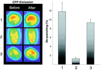
FRET analysis of the GABPαβ heterodimer after acceptor photobleaching. C2C12 cells were transfected with equal amounts of vectors encoding EYFP- and ECFP-GABP fusion proteins. Images were taken before and after the bleach pulse, using both the CFP and FRET filter sets. Left panels, after the cells were differentiated into myotubes, representative images (before and after photobleaching) of the following GABP wild-type and mutant protein combinations were obtained with the ECFP filter set (λ = 480 nm): 1, EYFP-N1-GABPα plus ECFP-C1-GABPβ; 2, EYFP-N1-GABPα(T280A) plus ECFP-C1-GABPβ; 3, EYFP-N1-GABPα plus ECFP-C1-GABPβ(S170A-T180A). Right panel, dequenching calculated as the increase in fluorescent intensity of the donor fluorophore ECFP after acceptor bleaching. Bars, 1, EYFP-N1-GABPα plus ECFP-C1-GABPβ; 2, EYFP-N1-GABPα(T280A) plus ECFP-C1-GABPβ; 3, EYFP-N1-GABPα plus ECFP-C1-GABPβ(S170A-T180A). Data are represented as the averages from ≥3 independent experiments with ≥3 individual cells. Error bars correspond to the standard errors of the means.

