FIG. 7.
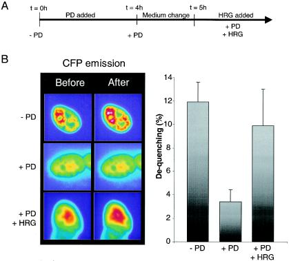
FRET analysis after phosphorylation modifications. (A) Stimulation protocol. At time zero, PD was added and incubated for 4 h. The cells were then washed and reincubated in differentiation medium for 1 h. The medium was again changed, and stimulation with HRG was initiated. (B) In order to determine the impact of phosphorylation on FRET efficiencies, dequenching experiments similar to those presented in Fig. 6 were performed. Left panel, images obtained before and after acceptor bleaching from C2C12 cells cotransfected with EYFP-N1-GABPα plusECFP-C1-GABPβ before (− PD) or after (+ PD) PD treatment or after medium change and reincubation with HRG (+PD + HRG). Right panel, dequenching calculations as described in the legend to Fig. 6.

