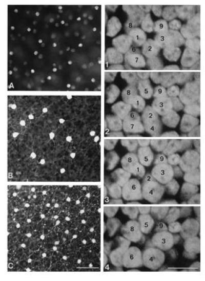
Figure 1.

(Left) Staining of specific amacrine cell populations. (A) Displaced starburst amacrine cells, stained by accumulation of DAPI. (B) Serotonin-accumulating amacrine cells in the inner nuclear layer, identified by immunohistochemistry. (C) AII amacrine cells, identified by immunohistochemistry. (Bar = 50 μm.) (Right) Serial horizontal sections (by confocal microscopy) through the inner nuclear layer. The nuclei were stained with ethidium homodimer. Cells were counted by following each cell from its beginning to end in the serial sections. (Bar = 10 μm.)
