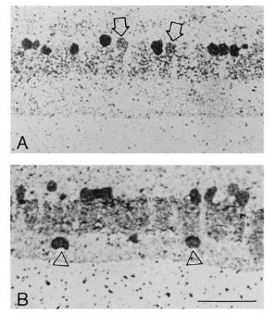
Figure 4.

Staining of etched semi-thin sections for endogenous GABA and glycine. Location 6 mm ventral to the optic nerve head. (A) Glycine-accumulating cells. (B) GABA-accumulating cells. In both, the ganglion cell layer is at the bottom. Arrows in A point to weakly labeled cells at the top of the inner nuclear layer, presumably glycine-accumulating bipolar cells. Triangles in B point to GABA-accumulating displaced amacrine cells, almost certainly starburst cells. They are shown as a positive control, since they are known to contain GABA (19–21). (They were not counted, because the analysis is concerned with the inner nuclear layer.)
