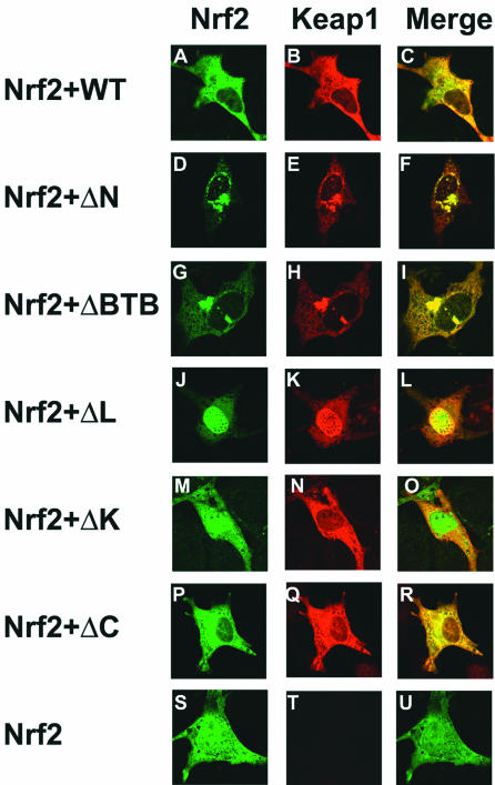
FIG. 2.
Cellular localization of the Nrf2 and Keap1 proteins. NIH 3T3 cells were cotransfected with an expression vector for HA-Nrf2 and expression vectors for either wild-type (WT) or mutant Keap1 proteins. The cellular localization of the Nrf2 and Keap1 proteins was determined by double-label indirect immunofluorescence with anti-HA (panels A, D, G, J, M, P, and S) or anti-Keap1 antibodies (panels B, E, H, K, N, Q, and T). Colocalization of the Nrf2 and Keap1 proteins is indicated by the presence of yellow in the merged images (panels C, F, I, L, O, R, and U).

