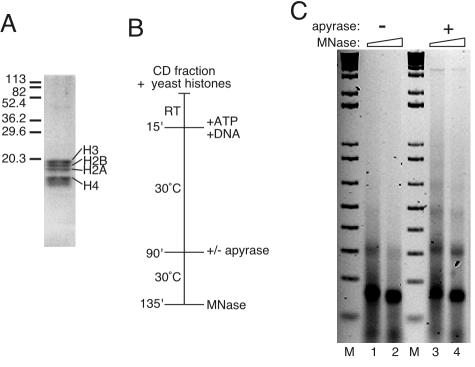
FIG. 4.
Yeast core histones form extensive nucleosomal arrays when ATP-dependent nucleosomal mobilization is inhibited. (A) Coomassie blue staining of yeast histones resolved by sodium dodecyl sulfate-polyacrylamide gel electrophoresis. The migration of molecular mass markers is indicated on the left in kilodaltons. (B) Flow diagram of reactions, which were performed by using the standard reaction cocktail. After 75 min, apyrase was added to half of the samples (lanes 3 and 4 in panel C), and all of the reactions were incubated for an additional 45 min. RT, room temperature; MNase, micrococcal nuclease. (C) Assembly products were assayed by micrococcal nuclease digestion, followed by agarose gel electrophoresis. The DNA was visualized with ethidium bromide. M, 1 kbp plus DNA ladder.

