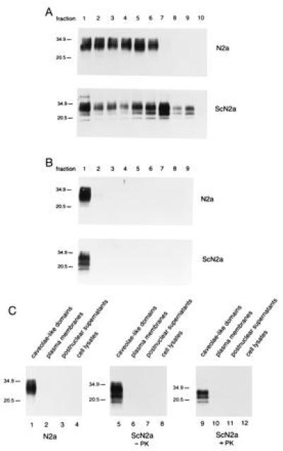
Figure 2.

PrPC and PrPSc are concentrated in CLDs isolated from N2a and ScN2a cells isolated by a detergent-free method. (A) Distribution of PrP in gradients after disruption of isolated plasma membranes by sonication and separation of CLDs from plasma membrane debris by flotation into OptiPrep gradients. Proteins precipitated from gradient fractions were subjected to immunoblot analysis using the anti-PrP polyclonal R073 rabbit antiserum (26). (B) Distribution of PrP after concentration of CLDs by step-gradient centrifugation. Immunoblot of proteins precipitated from step-gradient fractions probed with the anti-PrP R073 antiserum. (C) Concentration of PrP in CLDs. Equal amounts of proteins derived from cell lysates, postnuclear supernatants, plasma membranes, and CLDs of N2a and ScN2a cells were subjected to immunoblot analysis with anti-PrP R073 antiserum.
